Bad Credit Payday Loans Get Quick Financial Help Effortlessly

People discolored with inadequate in addition to a bad credit score ratings end up finding on their own inside problems even if they're trying to find exterior financial support to be able to deal with further costs in the middle of the particular 30 days. When they search for high street loan companies and make an application for the lending options right now there after that using loans are declined because of getting high risk elements. The reason being getting lending options inside traditional loan marketplaces is very difficult. However, you do not need to be able to damage the coronary heart given that poor credit payday advances are usually planned and also accessible to match your economic needs even with having poor credit records.

A bad credit score payday loans are especially made for those who find themselves poor credit borrowers and therefore are advanced to get a extremely short period of time period of time. In order to cope with the pressing or perhaps unforeseen needs, you can get their hands on the fund ranging from $100 to be able to $1500 for your amount of 2 to 4 weeks. The particular finance usually receives transited straight into your own lively bank account of the customer inside a matter of a day of right after applying. Interest of these loans is higher as compared to the standard lending options yet a planned out on the web loan general market trends can assist you get the better monetary deal.

Through the assistance of a bad credit score payday advances it is possible to pay back medical bills, small vacation to country side with family, abrupt automobile fixes, food store bills, space leases, paying down bank card fees, paying for childs school charges and much more. These financing options also help you to seize cash to handle their requirements and requirements.

Cataract - types, causes and treatment


What is cataract?

Cataract is one of the eye ailments that need screening of lens and proper medical attention. Cataract is a disease of eye lens that results in visual disturbances. In eyes, lens act as a focusing lens that pass the light to inner parts of eye. Cataract causes progressive but painless, lens opacity similar to clouding due to pathological changes in lens crystalline protein. Depending on the type and duration of disease development, it may affect the passage of light through the lens. In age-related cataract, disease process starts after age of 60 that proceeds gradually. Patients with pre-disposing factors are considered as ‘at-risk’ individuals. Usually, cataract affects one eye but it is possible to develop in another eye.

Types and causes

There are four types of cataract of which, age-related (senile) cataract is the common type. Other types of cataract are congenital, secondary and traumatic cataract. Each of these types may cause due to various factors and predispositions. In some cases, the exact cause remains unknown.

  • Senile cataract – Senile cataract causes as a result of free radicals-mediated inflammation and denaturation of lens protein due to aging process. The opacity may be partial or complete depending on type of cataract. In the initial stages, senile cataract can be treated by surgical and/or medical interventions and hence complications can be prevented. 


  • Congenital cataract - Occurs at birth due due to various factors such as underlying genetic disorders including Turner’s syndrome and galactosemia, infections such as congenital syphilis and rubella, familial history of cataract and other birth defects. 


  • Secondary cataract – Occurs as a result of eye surgery for other eye diseases, certain medicines such as water-pills, steroids and haloperidol group of drugs, iodine deficiency, increased blood pressure, smoking habits, metabolic disorders such as diabetes and thyroid dysfunctions, rheumatoid arthritis, alcohol consumption, radiation rays such as X-rays, prolonged and direct exposure to UV and infrared rays, atopic allergic reactions, infections such as leprosy and toxoplasmosis. 
  • Trauma-induced cataract – Occurs as a result of previous accidental eye injury and/or lens damage by mechanical or shrapnel injuries.

Symptoms

The initial stages of disease are usually asymptomatic in nature. As disease progresses, the patient may experience abnormal visual symptoms such as opaque/ cloudy or filmy vision with halos around light objects, blurred-double vision, lack of clear vision, abnormal sensitivity to sunlight and bright light objects, impaired night vision and inability to distinguish different color shades. Most of the patients experience short-sightedness and vision changes that needs frequent visit to opticians and changing of spectacles.

Diagnosis

Cataract can be differentially diagnosed by:

  • Slit lamp examination
  • Routine eye examination by using Snellen chart for vision acuity
  • Eye dilation examination
  • Tonometry test to rule out co-existing disorder like glaucoma.

What happen if left untreated?

Cataract can cause vision changes, inability to distinguish some colours like blue, phacomorphic glaucoma and irrevocable vision loss, if left untreated. Presence of co-existing diseases such as glaucoma or macular degeneration may worsen symptoms of cataract. Hypermature cataract is a serious complication that may occur in end-stages of untreated cataract which can be characterized by rupture of lens environment and leaking of internal fluids.

Treatment

Cataract in the initial stages can be treated without surgical procedures. Some of them are: Better eye glasses with eye drops that contain azapentacene and protective sunglasses for outdoor activities can be helpful. These non-pharmacological interventions can slow-down the progression of disease process but surgery is the only treatment, if symptoms get worsen. Surgical procedure involves removal of cataract by a process of phacoemulsification, followed by aspiration of lens fluid. The subsequent process is replacement of lens with intra-ocular lens (IOL) which may be monofocal or multi-focal depending on the requirement. Surgeons prefer fewer incisions to prevent after-surgery complications and inconvenient symptoms. After surgery, antibiotics are prescribed to prevent surgery-induced infection and some patient may need eyeglasses for proper vision. These surgical procedures are common for children as well as elderly patients.
How you can prevent cataract?

Prevention is better than cure – lifestyle modification by quitting smoking and alcohol habits, protective sunglasses to avoid exposure to UV, limiting edible substance that contain lactose such as milk and other dairy products, proper treatment and medical management of metabolic disorders and infectious diseases, intake of green vegetable and fruits that are rich in anti-oxidants, nutritional supplements rich in Vitamin E & C can be helpful to prevent incidence of this eye disorder in ‘at-risk’ individuals and also in aged individuals.

Reference: Zigler JS Jr, Datiles MB III (2011). Pathogenesis of cataracts. In: Tasman W, Jaeger EA (Editors). Duane's Ophthalmology. 15thEdition. Philadelphia, Pa: Lippincott Williams & Wilkins; 72B.

Costochondritis diagnosis

Costochondritis diagnosis

How is the diagnosis?
The diagnosis is made based on typical clinical history, findings on medical examination and the exclusion of other diseases.

The main symptom is pain in the chest wall of varying intensity and is often described as sharp, aching or pressure-like. The pain is eventually aggravated by movements of the upper body by deep breathing and physical exertion. Prior prolonged cough, severe bodily stress or physical activities that are charged to the arms, are common. Although costochondral joints often are inflamed, then each of the seven transitions Costochondral be inflamed. Pain may exist in several joints, but often state only on one side.

Costochondritis diagnosis
Pain that is recreated when the doctor pressing against the Costochondral parties in the chest wall, suggesting costochondritis. Movements in the arm on the sick side will often trigger pain one. It is particularly important to rule out that the pain stems from the heart Arch Intern Med 1994; 154: 2466-9. PubMed, 10 Miller CD, Lind sells CJ, Khandelwal S, et al ., an ECG heard so immediately. Sometimes the doctor will refer to X-ray of the chest.

Costochondritis treatments

What is the treatment?
The condition is referred to be self-limiting. Any treatment aims to relieve pain. Current assets is as paracetamol and / or NSAIDs. Activities that trigger pain, should be limited. For example, cough suppressants be useful for troublesome cough.

Costochondritis causes

Costochondritis causes


What causes Costochondritis?
By Costochondritis, there is an inflammation (inflammation) in several of his ribs bruskvev. The reason why there is inflammation, we do not know for sure. Many of the patients with Costochondritis can talk about prolonged cough, that they have recently made a significant physical effort (lift) or bodily activities that are charged to the arms.

What is the prognosis?
Almost everyone gets rid of pain. The course is variable and can last from weeks to months, but the condition is usually over in less than a year.
Costochondritis causes

Costochondritis

  • Costochondritis is an inflammation of several of the bruskete transitions between ribs and sternum.
  • It is a harmless condition that goes by itself.
  • The main symptom is pain in the chest wall of varying intensity and is described as sharp, aching or pressure-like.
  • The pain is located behind the sternum, often only on one side.

Chest wall pain costochondritis

Chest wall pain costochondritis


How is the chest wall built?
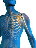
Ribs (costa) consist of bones caused by the disks and to whip forward and slightly downward. The upper seven ribs attach to the breastbone (sternum). In the transition between the ribs and the sternum is a cartilaginous portion that forms an elastic bridge - a sort of joint (costochondral paragraph) between the bony part of the ribs and sternum. Ribs 8-10 attach front the bruskete part of the overlying rib. The lower two ribs end blindly and is not attached to any other structures.
Chest wall pain costochondritis


Ribs move when we breathe, when we twist or bend the body and the movements of the arms.

Acute pulmonary edema

Acute pulmonary edema


What is an acute pulmonary edema?
Your lungs contain millions of small, elastic lung sacs called alveoli. With every breath, you take, transfer oxygen from the alveoli to the blood and the other way separated carbon dioxide, a waste product from burning out from the blood into exhaled. Normal takes place this exchange of oxygen and carbon dioxide without any problems. However, it sometimes causes increased pressure in the arteries in your lungs that liquid seeps out, fill the alveoli and prevent them from taking up oxygen - a condition called pulmonary edema.

In most cases, it is a heart disease that causes pulmonary edema. However, fluid can accumulate in your lungs for other reasons, such as pneumonia, certain toxins or drugs, or if you have acute altitude sickness.
Acute pulmonary edema

Acute pulmonary edema is a medical emergency and requires immediate treatment. Although pulmonic edema, sometimes leading to death, so the outlook is often good if you get quick and appropriate treatment for pulmonary edema and for the underlying disease.

What is costochondritis

What is costochondritis


Chest pain is something most people experience from time to time. Fortunately, the explanation in most cases harmless. A frequent cause of chest pain is inflammation of the cartilage between the ribs and the transitions' sternum.

What is costochondritis?
Costa means ribs, while chondritis means inflammation of the cartilage. Other designations are costochondritis costosternal syndrome, or anterior parasternal condrodyni chest wall syndrome Costochondritis is a self-limiting condition in which there is inflammation in several of the bruskete transitions between ribs and sternum Proulx AM, Zryd TW. . The condition produces pain fortil the chest wall, and it is often confused with Tietze syndrome.
What is costochondritis

Costochondritis is a condition that can occur in children as well as in adults. It is a common diagnosis in adults with acute chest pain, and the condition is stated to be the explanation for chest pain in 10-30% of these patients.

Free medical care

Free medical care


A MEASURE "TIMELY" AND "COURAGEOUS"

The workshop on health financing, opened Thursday at the headquarters, was an opportunity for the Department of Health and the Fight against AIDS to publish the first results of the measurement of free medical orderly.

Thus, after the first wave of action that took place from April 16 to May 31, a number of areas have been borrowed in order to assess the effect of the extent of exemption from payment of the use of health services, as well as the cost of health services.

The results of the study presented by Dr. Raymond, Deputy Director General of Health, report that the extent of exemption of payment of care determined by the Ivorian government "was timely and justified by the precarious situation of generalized population created by the post-election crisis."

Furthermore, this decision described as "courageous" by the country representative, was welcomed by the population, has demonstrated the application of health services by institutions of mass participation care throughout the period concerned.

However, he lamented; the provision of care has suffered quantity and quality of the low satisfaction of drug needs and strategic inputs hospitals. Hence the present shortage in several institutions.
Free medical care

The post-election crisis, from December 2010 to April 2011, led the Ivorian government to decide on the exemption from payment of care from April 16 to May 31, 2011 by users of public health facilities, para public and community contracted. However, the scale of need has led the President of the Republic to declare free health care "until further notice."

Kawasaki disease prognosis

Kawasaki disease prognosis


What is the prognosis?
The condition is most benign and self-limiting, but it can therefore, rarely cause serious heart complications. Without proper treatment occurs aneurysms in 20-25% of cases and myocardial infarction in 1-2%.

At present, treatment with immunoglobulin and ASA, reduced the percentage of aneurysms below 5%.

Kawasaki Syndrome Facts

Febrile illness in children with rash, mucosal changes and lymph node enlargement.

The condition is self-limiting, but untreated it develops in 20-25% bulge in the blood vessels, especially coronary artery to the heart.

The disease affects children aged three months to 12 years, 85% are under five years a . Isolated cases in adults are described.

There are a number of factors that contribute to disease. Indications are that there is an infection that triggers the disease.

Symptoms can vary. It Must have a fever for more than five days, conjunctivitis, rash, changes in the oral mucosa, changes in the hands and feet, lack of effect of antibiotics, lymphadenopathy in the neck (often lacking).
Kawasaki disease prognosis

Since diagnosis is difficult to ask, many will get antibiotics without help.

The dilemma is the risk of complications and serious sequela.

Kawasaki syndrome

Kawasaki syndrome


Follow-up
Plan for follow-up depends on several factors. These include the age when one becomes ill, the effect of treatment and the size of any ballooning of the arteries. The current guidelines released in 2004.

Doctors found no or small, transient changes in a coronary arteries, there is no need for special attention beyond the medical checkups every 3 to five years. These controls should include an assessment of the overall risk of cardiovascular disease and counseling to reduce this. There is no need for special inspections or monitoring by the cardiologist.

Doctors found a small change in the coronary arteries; the child should continue with medication (ASA) until the changes have gone back. There are no restrictions on physical activity except for the first 6-8 weeks in children under 11 years. Children over 11 years of age must receive individual advice on physical activity depending on the findings on cardiac examinations. After the initial phase of illness, it is recommended annual follow-up by the cardiologist.
Kawasaki syndrome

In the case of larger or more ballooning of the arteries, put the child on long-term treatment with blood thinners. Contact Sports discouraged until the cardiologist may find that the changes have gone back. It is recommended follow-up twice a year by the cardiologist. The same recommendations apply if it is shown that the arteries are clogged.

Kawasaki disease treatment

Kawasaki disease treatment


Treatment
Since the condition is reminiscent of an infection and the diagnosis is difficult to ask, many will have received antibiotics without effect. Regardless of the disease in its acute phase transition by itself.

The dilemma is the risk of complications and serious sequela. If the diagnosis is made, doctors will therefore provide direct immunoglobulin in the blood to reduce the harmful effects of the disease. This treatment provides rapid improvement in the condition with fever of freedom within one to two days. The treatment also reduces the risk of later heart disease. There will also be given acetylsalicylic acid (ASA, ex. Dispril) in a few weeks.

Kawasaki disease diagnosis

How is the diagnosis?
Diagnosis is made primarily on the medical history, and finds the doctor do when the child is physically examined. Blood tests do not contribute to make any definite diagnosis, but may be instructive. X-ray of the lungs can sometimes show mild pneumonia-like changes.
Kawasaki disease treatment

If doctors suspect the diagnosis, the echocardiogram show changes in the heart that support the diagnosis.

Kawasaki syndrome symptoms

Kawasaki syndrome symptoms


What are the symptoms of Kawasaki syndrome?

Symptoms and signs develop over the first 10 days, and the disease goes back by itself in most children, even without treatment. Symptoms can vary, but most have:


  • Fever for more than five days without explanation
  • Conjunctivitis
  • Rash - are almost always.
  • Lymph node enlargement in the neck - often lack.
  • Changes in the oral mucosa
  • Changes at the hands and feet
  • Lack of effect of antibiotic

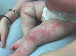
A number of other symptoms may also occur in connection with the disease: eg. irritability, joint pain, diarrhea, abdominal pain and ear pain. The changes in the oral cavity consist of redness, cracking and / or crust formation on the lips and strawberry tongue. The changes in hands and feet made of thickened skin, swelling and redness of the palms and soles, desquamation after approx. two weeks.
Kawasaki syndrome symptoms

Symptoms and signs develop over the first 10 days, and the disease goes back by itself in most children, even without treatment.

Kawasaki syndrome causes

Kawasaki syndrome causes


What causes Kawasaki Syndrome?
The cause is unknown. Probably, there are a number of factors that contribute to disease, but there are indications that there is an infection.

However, they have not been able to detect any virus or microbe.

In the acute phase developed inflammation of the wall of the coronary arteries (coronary vasculitis).

This can lead to a moderate expansion of the coronary artery.
Kawasaki syndrome causes

The lesions may persist from a few weeks to over a year after the disease onset.

What is Kawasaki Syndrome

What is Kawasaki Syndrome


What is Kawasaki Syndrome?

It is a febrile illness in children with rash, mucosal changes and lymph node enlargement. There is inflammation in the body's blood vessels (vasculitis). The condition is self-limiting and resolves by itself. Although the condition is by itself, one can get without late effects of treatment. The development of ballooning (aneurysm) of a coronary arteries (coronary arteries) in 20-25% if you do not receive treatment. In time, these sinuses interspersed cause serious complications. The disease was first described in Japan in the 1960s.

The disease affects children aged from three months to 12 years, 85% are under five years a . Isolated cases in adults are described. Sweden has found an annual incidence of 6.2 cases per 100,000 under five. The incidence of the disease varies throughout the year, with peaks in winter and spring.
What is Kawasaki Syndrome

The syndrome is most commonly in Asians, a little less frequently among blacks and lowest among whites. The incidence in the West is increasing.

Dilated cardiomyopathy prognosis

Dilated cardiomyopathy prognosis


Pregnancy-related cardiomyopathy

What is the prognosis?
Unlike the second cardiomyopathies are more people with cardiomyopathy periparital healthy by themselves, and they recover heart function. In a study from South Africa, 15% died, while 23% had recovered normal cardiac function after six months of treatment. Improvement, however, was observed both in the 2 and three year after the diagnosis was made, so that improvement phase is longer than the first 6-12 months.

A new pregnancy in a woman who has had periparital cardiomyopathy, provides a clearly increased risk of fresh heart failure. The greatest risk is for those who still have a remnant of heart failure after previous pregnancies.
Dilated cardiomyopathy prognosis

One study showed that half of those who became pregnant again, went through the next pregnancy, unless there were signs of new disease and heart failure.

Treatment cardiomyopathy

Treatment cardiomyopathy


Pregnancy-related cardiomyopathy

What is the treatment?
Since we do not know the cause of the disease, then we cannot give medicine to treat the disease itself. The treatment, therefore, be directed towards heart failure. Medications such as ACE inhibitors, beta blockers and digitalis use. Typical turnaround time is at least one year. In severe cases, it may be necessary with heart transplantation.

Otherwise you should be careful about using too much salt. You should also limit alcohol intake. It is important to try to be physically active.

How is the diagnosis?

The diagnosis involves the exclusion of other possible explanations for the condition. The main investigation is ultrasound of the heart ( echocardiography ).
Treatment cardiomyopathy

This study can provide both descriptive pictures of the extent of heart muscle damage, and it can quantify the degree of eventual heart failure.

Signs and symptoms of cardiomyopathy

Signs and symptoms of cardiomyopathy


Pregnancy-related cardiomyopathy

What are the symptoms and signs, disease?
The condition usually presents with symptoms and signs of heart failure. There is an increased amount of water retention (swollen legs), breathing difficulty on exertion, breathing difficulty when lying but not when you sit up, nocturnal attacks of breathing difficulty, persistent cough, discomfort from the stomach, heart palpitations.

Early symptoms and signs are often overlooked and can be camouflaged by the pregnancy; they are perceived as normal pregnancy signs.


In some cases, thrombosis with obstruction of blood vessels (arteries) in the legs (trunde gangrene), brain (stroke), in the intestine (threatening gangrene), in the heart (heart attack). Such complications can be life threatening.

Causes of cardiomyopathy

Causes of cardiomyopathy


Pregnancy-related cardiomyopathy

What causes peripartum cardiomyopathy?
The cause and the underlying disease mechanisms of peripartum cardiomyopathy is unknown, and many theories have been proposed. Nutritional disorders, autoimmune disease, inflammatory disease, viruses, blood cells from the fetus that enters the mother's blood as well as other theories have not led to any conclusion about the cause or progression of disease.

We are also uncertain about what might predispose to this disease. It is suggested that the following factors may increase the risk: high maternal age, number of previous births, African origin, pre-eclampsia, high blood pressure, use of rihemmende agent and twins.
Causes of cardiomyopathy.jpg

Despite this, we know about. 1/3 of the cases occur among young nulliparous.

Pregnancy related cardiomyopathy

Pregnancy related cardiomyopathy


What is pregnancy-related cardiomyopathy?

Pregnancy-related cardiomyopathy (peripartum cardiomyopathy) is a condition of unknown cause in which early signs of heart failure and symptoms of heart failure occur between the last month of pregnancy and the first five months after birth. However, there are reports of similar disease pictures earlier in pregnancy, cases may be due to this condition.

Cardiomyopathy means that the disease attacks the muscles of the heart. Peripartum means surrounding the birth. The diagnosis of peripartum cardiomyopathy is made by excluding other causes of cardiomyopathy.
Pregnancy related cardiomyopathy

A heart that fails in this condition will cause the heart expands - dilated - is greater. It increases the risk of blood clots can form in the heart. Such blood clots can break loose, float in the blood and clog blood vessels elsewhere in the body.

Peripartum cardiomyopathy is a relatively rare disease. There are no reliable prevalence data, and the numbers that exist, differ widely. In the U.S., it is suggested that there are approx. 1 case per 4000 births.

Treatment for hypertrophy

Treatment for hypertrophy


Preventive treatment

Young people with known hypertrophic cardiomyopathy should be advised not to expose themselves to excessive exertion, even if their condition is asymptomatic.

They should therefore, not participate in competitive sports with high standards of fitness. Most, however, participate in recreational activities that cause only mild to moderate physical strain.

How are long-term prospects?

Many patients may be asymptomatic throughout life and have a normal life. Among those who will develop these symptoms usually slowly. Over time, it can develop heart failure or angina pectoris.
Treatment for hypertrophy

Sudden death can occur, but modern treatment with the defibrillator and cardiac surgery aims to reduce the risk of such events.

Treatment for hypertrophic cardiomyopathy

Treatment for hypertrophic cardiomyopathy


What is the treatment?

The goal of treatment is to reduce symptoms and possibly prevent complications, particularly sudden death.

People without symptoms, and where there is no information on family cases of unexpected death at a young age, should not be treated medically, but is followed by a heart specialist.

Drug therapy is the primary treatment if the patient has symptoms. In complicated cases where medicines are working poor, both pacemaker therapy and surgical treatment are appropriate.
Treatment for hypertrophic cardiomyopathy

In some cases, a defibrillator implanted under the skin and in extreme cases, heart transplantation may be required.

Hypertrophic cardiomyopathy diagnosis

Hypertrophic cardiomyopathy diagnosis


How is the diagnosis?
Medical history and any ECG changes suggestive of cardiac disease. In the further investigation is echocardiography, ultrasound of the heart, the main survey. Here, the doctor can see the thickening of the heart wall, and any abnormal pressure differences in the heart can be measured. When your doctor does not find evidence of another disease that could explain the enlarged heart, we can conclude that there is hypertrophic cardiomyopathy.

Other investigations that may do are exercise-test and 24-hour heart rate monitoring. Sometimes also made ​​MRI of the heart. Genetic tests are of little use.

It is recommended that first-degree relatives (parents / siblings / children) examined the diagnosis in a person. Although family members do not have symptoms or signs of disease, it may still be changes in the heart.
Hypertrophic cardiomyopathy diagnosis

It is usual to follow the relatives of controls (ECG and echocardiography) every other year in young adulthood and every five years in adulthood.

Signs and symptoms of hypertrophic cardiomyopathy

Signs and symptoms of hypertrophic cardiomyopathy


Symptoms and signs
Patients with hypertrophic heart disease, for many years without symptoms of heart disease. If symptoms recur, the most common breathing difficulty and chest pain on exertion ( angina ).

Some brand's palpitations or anxious heart. Some shy of dizziness and fainting, often triggered by exertion.

Examination by a doctor may not be very wrong to find in the early stages. However, there may be a murmur over the heart.

ECG , however, usually not normal. It often shows signs of altered flow distribution in the heart (conduction disorder) and various degrees of thickened muscle of the left ventricle.
Signs and symptoms of hypertrophic cardiomyopathy

Sometimes one can find atrial fibrillation or other arrhythmias.

Cause of hypertrophic cardiomyopathy

Cause of hypertrophic cardiomyopathy


What is the cause of hypertrophic cardiomyopathy?

It Must have a family history of known cases of heart disease and sudden cardiac death. The condition is hereditary and is caused by mutation (error) in one or more genes. Gene's defect leads in turn to the muscle cells of the heart grows too much.

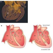
The changes in the heart can vary from person to person. Many small changes and have no complaints. Typically, the wall of the left ventricle (ventricle), which is thickened (hypertrophic). At 20-30% will be the end of the left ventricle cramped, making it difficult for the heart to pump blood out into the wide circulation. Furthermore, anterior chamber (atria) may eventually be enlarged.
Cause of hypertrophic cardiomyopathy

The changes in heart muscles and the atria predisposing to arrhythmias in the heart. In some cases, these arrhythmias may be severe and lead to cardiac arrest.

What is hypertrophic cardiomyopathy

What is hypertrophic cardiomyopathy


Enlarged heart - hypertrophic cardiomyopathy

Cardiomyopathy means disease of the heart muscle. Hypertrophy means enlargement. Hypertrophic cardiomyopathy is a disease where the heart muscle is enlarged, and were in some cases lead to symptoms due to impaired cardiac function.

Typically, an enlarged heart due to underlying disease such as congenital valvular abnormalities or high blood pressure. However, the condition hypertrophic cardiomyopathy is no such underlying disease. The disease is hereditary and is due to changes in genes (mutations) that can be transmitted to new generations.
What is hypertrophic cardiomyopathy

Hypertrophic cardiomyopathy occurs in approximately. 1 of 500-1000 adults. The disease is present from childhood, but rarely produces symptoms until puberty. Often it is discovered wounded in youth or adulthood. The condition is the most frequent cause of sudden cardiac death among young people and athletes.

Hyperlipidemia treatment guidelines

How is hyperlipidemia?


Treatment goals
If it is necessary to treat, apply the following treatment goals for fats in the blood:

  • Total cholesterol less than 5.0 (mmol / L)
  • Relationship between the values ​​for total cholesterol and HDL cholesterol (good cholesterol) should be less than 4
  • LDL cholesterol should be lower than 3.0 (mmol / L)
  • Triglycerides should be less than 2.0 (mmol / L)
  • HDL cholesterol should be higher than 1.0 (mmol / L)

General treatment
Total risks for heart disease are essential for the gain of fat-lowering treatment. A slightly elevated cholesterol is isolation tungveiende no reason for treatment; it is the sum of risk factors is basic. That is, the more of the factor's hyperlipidemia, hypertension, diabetes, genetic predisposition, smoking, obesity, lack of exercise that is, the more important it is to manage to avoid the development of cardiovascular disease.

All treatments start with a change of diet. The next step in treatment is the use of fat-lowering drugs, called statins. In some cases, it may also be appropriate to use triglyceride funds.

Dieting recommended for obesity. Smoking is a major risk factor for vascular disease of the heart and smoking cessation is therefore, important. Regular exercise improves the composition of fats in the blood. It is often necessary to get a permanent weight loss. The goal is at least 30 minutes of exercise 2-3 times a week.
Hyperlipidemia treatment guidelines

The isolated elevated triglycerides recommended low alcohol intake, caution with coffee, weight loss in overweight people.

What if you have elevated cholesterol, but otherwise no other risk factors?
  • When cholesterol levels between 6.5 to 7.9 and LDL-cholesterol of 5.0, or a relationship between LDL and HDL over 5, the non-drug therapy
  • Cholesterol of 8.0 is treated first with drugs.
  • If LDL-cholesterol continues to be above 5.0, or the relationship between LDL and HDL continues to be above 5 at follow-up, drug therapy should be considered.
  • Women are not treated medically until after they have ceased to menstruate.

Self-Treatment
The restructuring of lifestyle should continue for at least six months before taking the drug therapy. The exceptions are patients with heart disease, which often starts with drug treatment and lifestyle change simultaneously.

Dietary advice
The amount of saturated fats in food is reduced to less than 10% of the total energy (calorie intake). The main sources of saturated fat in the diet are meat, meat products (15-20%), margarine and milk products (40%). Limit your total fat intake to 30% of energi-/kaloriinntaket Cover 50% of energy intake from carbohydrates - cereals, fruits and vegetables. Increase your intake of fatty fish, soft margarines and olive oil. Have a moderate alcohol consumption. A intermediate consumption of nuts, 50-100 g daily, can reduce cholesterol levels by approx. 0.3 mmol / l.

Pharmacotherapy
Cholesterol-lowering medications given to patients without known heart disease if they are at high risk of heart attack.

In patients with known cardiac disease or symptomatic disease that confined Halske, stroke, or poor blood circulation, offered cholesterol-lowering drugs to patients with cholesterol levels below 5.5. The decision on the treatment a physician and patient assess the benefits, and the side effects and disadvantages of treatment can have. Treatment is often up to 65-75 years of age. The benefit in the elderly is under discussion.

Familial hypercholesterolemia

It is reasonable to believe that adult family members with LDL-cholesterol above 5.5 have familial hypercholesterolemia. Treatment Start from 18 years of age or earlier are implemented at high risk for vascular disease of the heart.

Isolated elevations in triglycerides

Consideration should be given medical treatment when the condition in the family, when the condition is combined with early heart disease and when triglycerides are over 10, because. Increased risk of inflammation of the pancreas.

Hyperlipidemia diagnosis

Hyperlipidemia diagnosis


How hyperlipidemia diagnosed?
It is important to clarify whether there is an accumulation of hyperlipidemia in the family. Are there relatives with elevated cholesterol? Are there cases of early heart disease (before age 60 years) in the family? User patient medication that may affect the lipid profile? If there are chronic diseases that may contribute to hyperlipidemia? Since hyperlipidemia is included as one of the several risk factors for heart disease, your doctor will also determine smoking habits, eating habits and alcohol intake.

The study sees the doctor for fat deposits in the eye area and around tendons. In order to assess the patient's overall risk of developing heart disease, your doctor will measure blood pressure, weight, listen to the throat, kidneys and groin arteries; feel for the pulse in the groin and lower legs and work out the relationship between livmål and hip measurement. This should be below 1.0 in males and below 0.9 in women.
Hyperlipidemia diagnosis

Measurement of fats in the blood is essential for diagnosis. This means the measurement of total cholesterol, HDL-cholesterol and triglycerides (fasting). The value for the dangerous part of the cholesterol (LDL cholesterol) can be calculated if one knows the values ​​mentioned above. Measurement of blood glucose is done to see if it could be diabetes. The cholesterol of 8 and isolated occurring elevations in triglycerides are taken in addition to glucose metabolism also hormones, liver test, kidney test and urine test strip examined for the presence of proteins (kidney damage). In many cases, your doctor will as well take a heart (ECG) to check for signs of damage of the heart.

How are long-term prospects?
Fat values ​​vary with age and gender. Fats appear to increase by 2-3 mmol / L from 20-30 years of age to 60-70 years of age. Long-term prospects depend on cholesterol levels, but primarily of the total risk of vascular disease of the heart. The complication of hyperlipidemia is heart disease.

Hyperlipidemia risk factors

Hyperlipidemia risk factors


Metabolic Syndrome
This is a condition with normal or slightly overhead total cholesterol. HDL cholesterol is reduced, and triglycerides are elevated. At the same time, these patients have elevated blood pressure and decreased tolerance for sugar, which means that many develop diabetes ( diabetes ). Another typical feature of these patients is obese around the abdomen.

Other issues
A number of diseases and medications can also contribute to the development of hyperlipidemia. It can apply to diabetes mellitus (diabetes), disorders of the thyroid gland ( hypothyroidism), kidney and liver disease, alcohol consumption, use of certain water pills; beta blockers may be unfavorable (heart medicine), cortisone drugs, and estrogens (female sex hormone).

What causes hyperlipidemia

What causes hyperlipidemia


Primary hyperlipidemia
The condition is due to inheritance of multiple genes that predispose to high cholesterol. In addition, the condition is often the lifestyle related. Cholesterol levels are moderately elevated and are usually around 6-8 mmol / L. The pattern of inheritance is such that hyperlipidemia occur scattered in the family. This means that not everyone in the family inherits the facility for the disease.

Familial hypercholesterolemia
This is called a dominant hereditary disease. This means that it is likely to inherit the condition, and the risk can be calculated as follows:
  • If one parent has a healthy and a diseased gene (called heterozygous) and the other parent is healthy, there are a 50% chance that a child inherits the disease.
  • If one parent has two diseased genes (called homozygous) and the other parent is healthy, all kids get sick.
  • If both parents have a diseased gene, there are a 75% chance that a child inherits the disease.
  • If both parents have two diseased genes, all kids get sick (and all will be homozygous)
What causes hyperlipidemia

The disease is characterized by the formation of grease balls in the eye area and along the tendons. The disease affects 1 in 400, but accounts for 5% of all cases of myocardial infarction. These individuals are therefore, at high risk for vascular disease of the heart. In the heterozygote, the complete cholesterol often be between 8 and 12 mmol / L. In homozygous, the total cholesterol eventually be above 20 mmol / L.

Familial combined hyperlipidemia.
The condition found in 1 of 50-100. It provides the same risk of disease in the heart as familial hypercholesterolemia. Total cholesterol is usually of 7-9 mmol / L, and triglycerides are above two mmol / L.

What is hyperlipidemia

What is hyperlipidemia?
Hyperlipidemia has high levels of fats in the blood. The term used for the increase of lipid cholesterol and triglycerides. Cholesterol can be measured as total cholesterol, HDL-cholesterol and LDL-cholesterol. HDL cholesterol is often termed "good cholesterol" because high HDL-cholesterol had been shown to protect against heart disease. Conversely, low HDL and high LDL cholesterol have an adverse effect on the heart.

Total Cholesterol
Limits of cholesterol (the concentration in blood is measured in mmol / L):

Less than 5, the desired level
5.0 to 6.4 implies easily elevated cholesterol
6.5 to 7.9 means moderately elevated cholesterol
Higher than 8.0 means expressed elevated cholesterol
Triglycerides
Triglyceride level higher than 2.3 mmol / L is considered elevation.
What is hyperlipidemia

Lipids as a risk factor
There is a clear association between cholesterol levels and risk of developing diseases of the heart. Elevated levels of triglycerides seem to be an independent risk factor for developing vascular disease of the heart, particularly in women.

Occurrence
In Norway, about 200,000 people a familial hypercholesterolemia in which some of the family has elevated cholesterol and where an inheritance pattern is related to several genes. Closely, 50,000 people have been familial combined hyperlipidemia in which both cholesterol and triglycerides are upraised. Approximately, 10,000 people have elevated cholesterol caused by a defect in a specific genetic material.

Pulmonary embolism prognosis

Pulmonary embolism prognosis


How are the course and prognosis?
In general, because the disease decided. Any worsening of cor pulmonale depends upon both on how early treatment starts and how effective this is. Complications are right-sided heart failure and right-sided heart attack.

Good treatment of grunnsykom well as the reduction of oxygen is important to avoid further forverrring of secondary pulmonary hypertension. The prognosis depends on the underlying disease. For example, patients with COPD and moderate respiratory prevent a 3-year mortality risk of 50% right-sided heart failure after occurred nine.

Pulmonary embolism prognosis
In patients who live many years with their basic disease, it may eventually develop cor pulmonale and right-sided heart failure If the patient develops right-sided heart failure, this could be treated on equal terms with left-sided heart failure.

Pulmonary treatments

Pulmonary treatments


What is the treatment?
Whatever the reason disease, low oxygen content in the blood exacerbating the patient's condition. The treatment involves so much to improve the patient's oxygen consumption though, for example, continuous oxygen therapy and to improve treatment of any disease do. If the patient develops right-sided heart failure, it must be treated with diuretics and cardiac tonic medicine (digitalis). Treatment of primary pulmonary hypertension is complex, controversial and potentially dangerous.

The most important measure is smoking cessation, for whatever reason disease. Snuff, chewing tobacco and nicotine products are alternatives if it is difficult to stop completely. Patients should avoid activities that aggravate breathlessness public. If there is excess, it is necessary to slow weight loss.
Pulmonary treatments

Different types of drugs may be necessary.

Diagnosis of pulmonary embolism

Diagnosis of pulmonary embolism


How is the diagnosis?
In most cases, you will find an underlying cause of pulmonary hypertension and cor pulmonale. Symptoms will vary with the trigger and will also be different in the acute and chronic course. The diagnosis of cor pulmonale requires the finding of an enlarged right ventricle or increased pressure in lungekarsengen.

Acute illness is illness characterized by severe heart failure. In chronic disease, illness characterized by the underlying disease.
Diagnosis of pulmonary embolism

ECG and lung function measurements may be useful in additional studies. The same applies to various types of image studies X-ray of the heart and lungs, echocardiography Schiller NB. , lung scintigraphy, CT and MRI, as well as measurements of blood gases. Cardiac catheterization is the most informative study. In some cases, it is necessary to take tissue samples of lung tissue.

Sign and symptoms of pulmonary embolism

Sign and symptoms of pulmonary embolism


What are the symptoms and signs' results in high blood pressure in the pulmonary circulation?

In most cases, you will have symptoms associated with a precipitating cause, eg. a blood clot or pneumonia. The most common symptoms - breathing difficulty striking the effort, fatigue and fainting - reflects the heart is unable to increase its pumping capacity during physical activity. For example, angina pectoris occurred despite normal blood vessels of the heart wall (coronary).

Diseases of the lung vessels
Acute blood clot to the lungs provides serious, severe breathing difficulties. Chronic diseases provide breathing difficulty, cough, chest pain and swelling in the legs.
Sign and symptoms of pulmonary embolism
Diseases that affect lung function.
These are diseases such as COPD, chronic asthma and musculo-squelettiques diseases and neurological disorders that inhibit lung function. Typical symptoms include difficulty breathing, if necessary. loshoste (COPD), frequent respiratory infections.

Pulmonary circulation vessels

Pulmonary circulation vessels


What causes high blood pressure in the pulmonary circulation?
In primary pulmonary hypertension, the cause is unknown, but the abnormal changes associated with blood vessels in the lungs. Secondary pulmonary hypertension is a complication of many diseases of the lungs, heart and chest cavity. Cor pulmonale is a consequence of prolonged high blood pressure in the pulmonary circulation in which the right ventricle has become enlarged. Right ventricle is relatively thin and have limited ability to appreciate, therefore, the right side of the heart could begin to fail if blood pressure in the pulmonary circulation are persistently high. It occurs when the right-sided heart failure.

Another contributing factor is that the blood becomes high rate of chronic lung disease in which the absorption of oxygen is reduced. The blood becomes "thicker / stickier," which further increases blood pressure in the pulmonary circulation.
Pulmonary circulation vessels

A number of conditions can cause secondary pulmonary hypertension and cor pulmonale, the most common are: COPD , chronic asthma , small and large blood clots , massive obesity with respiratory problems, lung disease due to intravenous drug abuse, congenital heart disease , pulmonary fibrosis , removed lung tissue during surgery, muscle weakness, significant spinal distortions, low metabolism , inflammation of blood vessels (vasculitis).

The pulmonary circulation

The pulmonary circulation


The prevalence of high blood pressure in pulmonary circulation?
Primary pulmonary hypertension is a very rare condition and the number of new cases per year reported to be ca. 1-2 per 1 million people. In childhood there is no gender difference, but after puberty, the prevalence was higher in women than men (1,7:1). The condition is most frequent in the age group 20 to 40 years.

Secondary pulmonary hypertension is more common but is underdiagnosed. The exact incidence is difficult to estimate because of the huge spread in the underlying causes.

The pulmonary circulation
Cor pulmonale arising as a result of underlying disease, are relatively common and is the third most common cardiovascular disease (by coronary artery disease and heart disease as a result of high blood pressure in the large circulation) . For example, half of patients with COPD, cor pulmonale. About 20% of patients hospitalized with heart failure, have the right ventricular failure with cor pulmonale.

What is pulmonary circulation

What is pulmonary circulation


What is high blood pressure in the pulmonary circulation?

Pulmonary circuit goes from the right ventricle and into the lungs via the pulmonary artery and are derived in lung tissue. Collecting Veins (veins) causes the blood back to the left atrium of the heart. The right ventricle which pumps blood into the pulmonary circulation. Conversely, the sinistral ventricle which pumps blood out into the wide circulation. Most cases of heart failure due to failure of the left ventricle, less frequently occurring failure of the right ventricle.

High blood pressure in the pulmonary circulation, pulmonary hypertension, is a complex issue characterized by nonspecific symptoms and signs and with many possible causes. The unknown cause is primary pulmonary hypertension. The known cause is secondary pulmonary hypertension.
What is pulmonary circulation

Prolonged high blood pressure in the pulmonary circulation leads to the development of a condition called cor pulmonale (cor = heart pulmonary = lungs that have to do). It is a condition in which the right ventricle is enlarged because this part of the heart has to pump out the blood with greater force to be able to drive the blood through the pulmonary circulation. Cor pulmonale can cause both acute and chronic. The sequela of cor pulmonale is failing right ventricle, a right-sided heart failure.

Untreated high blood pressure in the pulmonary circulation, for whatever reason, will result in right-sided heart failure.

Heart disease long term effects

Heart disease long term effects


How are long-term prospects?
Heart failure is a serious condition in which it has already caused permanent changes in the heart. Medications used are of great help, and contributes significantly to alleviate symptoms and prolong life.

How good is the prognosis will depend on how severe the heart failure is?

Illustrations
Thorax, normally
Heart failure, severe
Heart disease long term effects

Facts
  • Condition in which the heart fails to pump blood around the body strong enough.
  • The most typical problems that arise, shortness of breath, decreased physical performance, lethargy, increased fatigue and decreased appetite.
  • Eventually, it may develop swelling in the legs and elsewhere in the body.
  • Different types of heart disease can cause heart failure, most commonly, post-myocardial infarction.
  • Years of high blood pressure can also be hard on the heart and cause heart failure.
  • Diabetes, infection, and alcohol can also weaken the heart.
  • Self-treatment combined with treatment in general practice or hospital.
  • The aim of treatment is to relieve symptoms, improve quality of life and increase life expectancy.

Disease condition of heart muscle

Disease condition of heart muscle


How is the condition?
Lighter grades of heart failure are treated in general practice. The pronounced heart failures are needed hospital treatment. The aim of treatment is to relieve symptoms, improve quality of life and increase life expectancy.

Self-Treatment
There are several things you can do:
Disease condition of heart muscle
  • Eat less salt. When you have a lower blood pressure, and make work easier for the heart. Salt bound fluid in the body, which increases the load on the heart. The total salt intake during the day should not exceed one teaspoon, corresponding to approx. 6 g of salt (NaCl). Limited intake of salt means cessation of all the extra salt added to the diet as in baking bread, potato cooking, etc.
  • Avoid large meals, eat less and do more frequent meals.
  • Avoid large amounts of alcohol.
  • Stump the smoke.
  • It may be helpful to lose weight if you are overweight.
  • Physical activity should be guided by symptoms. Can you regularly exercise a little; it is useful? However, you should not charge more than your body allows.
Pharmacotherapy
There are many drugs that are effective in heart failure. The major drug groups are:
  • ACE inhibitors work by widening blood vessels so that blood pressure goes down, and the load on the heart is reduced. This type of drug is today due to treatment in the vast majority of heart failure.
  • Beta blockers have the heart to beat slower, they lower blood pressure and strengthens the heart. Furthermore, this type of drug used by most people.
  • Diuretic reduces fluid in the body and thus the strain on the heart. It is part preferably in a form of triple therapy with ACE inhibitor and beta blocker.
  • Digitalis strengthens the heart. These were widely used before, but used less these days.
  • There are also other products that are beneficial in heart failure, and a host of new treatments is being developed and tested.
Blood thinning medications are used in some cases, especially if you have atrial fibrillation. Rhythm Stabilizing drugs are also used if you have the tendency to an irregular heartbeat.

Another treatment
For some it may be appropriate with surgery. This is particularly the flap failure. Among patients with a tendency to serious arrhythmias may be necessary to operate a pacemaker or electric equipment (ICD). Very rarely do a heart transplant.

How to diagnose heart failure

How the condition diagnosed?


The diagnosis will usually be suspected based on the typical symptoms are present. These are mentioned above. It will have blood tests to identify disease, and radiography of the chest is also relevant to distinguish heart disease from lung disease with similar symptoms. ECG may show signs of heart disease.

How to diagnose heart failure

Is there any doubt about the diagnosis, an ultrasound of the heart ( echocardiography ) to decide with certainty, whether there is heart failure or not? Occasionally, it may be necessary to do another research to clarify the cause of heart failure and the extent of the damage to the heart.

Causes Of Heart Failure

Causes Of Heart Failure


What causes heart failure?
When we are in motion, increases metabolism throughout the body. The different body parts need then more blood to get enough oxygen and nourishment. A healthy heart can increase their activity so that blood circulation is satisfactory. A diseased heart is not capable of this, and the condition known as congestive heart failure.

Different types of heart disease can cause heart failure, but the most common is that it occurs after myocardial infarction. A portion of the heart muscle is dead then, and it is replaced by a rigid scar tissue. This reduces the hearts pumping ability. Years of high blood pressure can also be hard on the heart and cause heart failure.
Causes Of Heart Failure

For the same reason, the failure of the heart valves ( aortic stenosis, aortic insufficiency, mitral stenosis, mitral regurgitation ) provide heart failure due to increased wear and tear of the muscle. There are also a number of conditions that weaken the heart (cardiomyopathy) such as diabetes, infection, alcohol.

Define Heart Failure

Define Heart Failure


What is heart failure?

Heart failure describes a condition where the heart fails to pump blood around the body strong enough. This means that each agency receives insufficient blood supply; the most typical problems that arise, the heavy breathing and impaired physical performance. For a mild degree of heart failure is marked by this first effort, while in severe heart failure have shortness of breath even at rest. You will also notice that breathing becomes heavier when you lie down, and it will be easier in an upright position. Eventually, it may develop swelling in the legs and elsewhere in the body. Other symptoms include weakness, increased fatigability, decreased appetite.
Define Heart Failure
Approximately, 50000-100000 have heart failure. Estimate that 10% over 75 years, congestive heart failure.

What is the prognosis

What is the prognosis


What is the prognosis?
Most case of acute pericarditis is a self-limiting, short-term course - often less than a week, and the prognosis is good. 10-30% of patients experience a relapse during the first months, but later decrease recurrence risk. Get (approx. 1%) developed constrictive pericarditis.
What is the prognosis
Bacterial pericarditis, which these days are a rare condition, may sometimes be life threatening. Furthermore, constrictive pericarditis can be life-threatening condition.

Treatment Pericarditis

Treatment Pericarditis


What is the treatment?
Patients with acute pericarditis usually observed at the hospital with ECG monitoring because it can be associated inflammation of the heart muscle (myocarditis) and thus the risk of an irregular heartbeat. Treatment is primarily symptomatic relief. Pain and fever treated with anti-inflammatory drugs (e.g. ibuprofen) and in most cases, colchicine tablets. Treatment usually continues for at least a week after symptoms are gone. Only exceptionally is cortisone treatment is required.
Treatment Pericarditis

If it accumulates a lot of fluid in pericardial, the patient must be monitored closely and evaluated for pericardial tapping, i.e., withdrawal of fluid from the cavity of the pericardium. Other rarer forms of pericarditis treated in relation to what is the underlying cause.

Symptoms Of Pericarditis

What are the symptoms of pericarditis?
Acute nonspecific pericarditis can occur without warning, or it can be assumed passed by a respiratory infection. Chest pain is the most frequent symptom and often occurs quite suddenly. The pain is usually intense, sharp and is located centrally or on the left chest. The pain sometimes radiates to the shoulders, neck, back, and possibly to the left arm, and can resemble the pain of acute myocardial infarction.

Typically, pericarditis merten is that it often gets worse when breathing deeply, coughing, swallowing or lying on your back. The pain is often relieved when sitting bent forward. The pain can last from quite a few hours to several days. Fever is often present, and there may be coughing and breathing difficulty.
Symptoms Of Pericarditis

In constrictive pericarditis, there is often a history of previous pericarditis, cardiac surgery or radiotherapy. Typical of this condition is increasing breathing difficulty, fatigue and weakness. The patient can tolerate little physical activity and are quickly out of breath with exertion.

What Causes Pericarditis

What Causes Pericarditis


What are the causes?
The main causes of acute pericarditis are viral infections (> 80%). Pericarditis can also be seen in tuberculosis or HIV infection (common in developing countries), or in connection with cancer. Other possible causes include kidney failure, and radiation damage.

In 5-10% of patients with acute myocardial infarction occurs a slight pericarditis after a few days. The condition is usually harmless, and it goes back by itself. As a result of modern treatment (thrombolysis), this pericarditis form becomes less frequent. Another type of pericarditis occurs later in throughout heart attack in 10-20% of infarct patients.
What Causes Pericarditis
The chronic constrictive form can occur as a result of radiotherapy (radiation to the tumor tissue in the chest cavity), heart surgery or previously performed viral pericarditis.