Costochondritis diagnosis

Costochondritis diagnosis

How is the diagnosis?
The diagnosis is made based on typical clinical history, findings on medical examination and the exclusion of other diseases.

The main symptom is pain in the chest wall of varying intensity and is often described as sharp, aching or pressure-like. The pain is eventually aggravated by movements of the upper body by deep breathing and physical exertion. Prior prolonged cough, severe bodily stress or physical activities that are charged to the arms, are common. Although costochondral joints often are inflamed, then each of the seven transitions Costochondral be inflamed. Pain may exist in several joints, but often state only on one side.

Costochondritis diagnosis
Pain that is recreated when the doctor pressing against the Costochondral parties in the chest wall, suggesting costochondritis. Movements in the arm on the sick side will often trigger pain one. It is particularly important to rule out that the pain stems from the heart Arch Intern Med 1994; 154: 2466-9. PubMed, 10 Miller CD, Lind sells CJ, Khandelwal S, et al ., an ECG heard so immediately. Sometimes the doctor will refer to X-ray of the chest.

Costochondritis treatments

What is the treatment?
The condition is referred to be self-limiting. Any treatment aims to relieve pain. Current assets is as paracetamol and / or NSAIDs. Activities that trigger pain, should be limited. For example, cough suppressants be useful for troublesome cough.

Costochondritis causes

Costochondritis causes


What causes Costochondritis?
By Costochondritis, there is an inflammation (inflammation) in several of his ribs bruskvev. The reason why there is inflammation, we do not know for sure. Many of the patients with Costochondritis can talk about prolonged cough, that they have recently made a significant physical effort (lift) or bodily activities that are charged to the arms.

What is the prognosis?
Almost everyone gets rid of pain. The course is variable and can last from weeks to months, but the condition is usually over in less than a year.
Costochondritis causes

Costochondritis

  • Costochondritis is an inflammation of several of the bruskete transitions between ribs and sternum.
  • It is a harmless condition that goes by itself.
  • The main symptom is pain in the chest wall of varying intensity and is described as sharp, aching or pressure-like.
  • The pain is located behind the sternum, often only on one side.

Chest wall pain costochondritis

Chest wall pain costochondritis


How is the chest wall built?
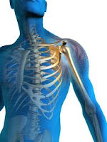
Ribs (costa) consist of bones caused by the disks and to whip forward and slightly downward. The upper seven ribs attach to the breastbone (sternum). In the transition between the ribs and the sternum is a cartilaginous portion that forms an elastic bridge - a sort of joint (costochondral paragraph) between the bony part of the ribs and sternum. Ribs 8-10 attach front the bruskete part of the overlying rib. The lower two ribs end blindly and is not attached to any other structures.
Chest wall pain costochondritis


Ribs move when we breathe, when we twist or bend the body and the movements of the arms.

Acute pulmonary edema

Acute pulmonary edema


What is an acute pulmonary edema?
Your lungs contain millions of small, elastic lung sacs called alveoli. With every breath, you take, transfer oxygen from the alveoli to the blood and the other way separated carbon dioxide, a waste product from burning out from the blood into exhaled. Normal takes place this exchange of oxygen and carbon dioxide without any problems. However, it sometimes causes increased pressure in the arteries in your lungs that liquid seeps out, fill the alveoli and prevent them from taking up oxygen - a condition called pulmonary edema.

In most cases, it is a heart disease that causes pulmonary edema. However, fluid can accumulate in your lungs for other reasons, such as pneumonia, certain toxins or drugs, or if you have acute altitude sickness.
Acute pulmonary edema

Acute pulmonary edema is a medical emergency and requires immediate treatment. Although pulmonic edema, sometimes leading to death, so the outlook is often good if you get quick and appropriate treatment for pulmonary edema and for the underlying disease.

What is costochondritis

What is costochondritis


Chest pain is something most people experience from time to time. Fortunately, the explanation in most cases harmless. A frequent cause of chest pain is inflammation of the cartilage between the ribs and the transitions' sternum.

What is costochondritis?
Costa means ribs, while chondritis means inflammation of the cartilage. Other designations are costochondritis costosternal syndrome, or anterior parasternal condrodyni chest wall syndrome Costochondritis is a self-limiting condition in which there is inflammation in several of the bruskete transitions between ribs and sternum Proulx AM, Zryd TW. . The condition produces pain fortil the chest wall, and it is often confused with Tietze syndrome.
What is costochondritis

Costochondritis is a condition that can occur in children as well as in adults. It is a common diagnosis in adults with acute chest pain, and the condition is stated to be the explanation for chest pain in 10-30% of these patients.

Free medical care

Free medical care


A MEASURE "TIMELY" AND "COURAGEOUS"

The workshop on health financing, opened Thursday at the headquarters, was an opportunity for the Department of Health and the Fight against AIDS to publish the first results of the measurement of free medical orderly.

Thus, after the first wave of action that took place from April 16 to May 31, a number of areas have been borrowed in order to assess the effect of the extent of exemption from payment of the use of health services, as well as the cost of health services.

The results of the study presented by Dr. Raymond, Deputy Director General of Health, report that the extent of exemption of payment of care determined by the Ivorian government "was timely and justified by the precarious situation of generalized population created by the post-election crisis."

Furthermore, this decision described as "courageous" by the country representative, was welcomed by the population, has demonstrated the application of health services by institutions of mass participation care throughout the period concerned.

However, he lamented; the provision of care has suffered quantity and quality of the low satisfaction of drug needs and strategic inputs hospitals. Hence the present shortage in several institutions.
Free medical care

The post-election crisis, from December 2010 to April 2011, led the Ivorian government to decide on the exemption from payment of care from April 16 to May 31, 2011 by users of public health facilities, para public and community contracted. However, the scale of need has led the President of the Republic to declare free health care "until further notice."

Kawasaki disease prognosis

Kawasaki disease prognosis


What is the prognosis?
The condition is most benign and self-limiting, but it can therefore, rarely cause serious heart complications. Without proper treatment occurs aneurysms in 20-25% of cases and myocardial infarction in 1-2%.

At present, treatment with immunoglobulin and ASA, reduced the percentage of aneurysms below 5%.

Kawasaki Syndrome Facts

Febrile illness in children with rash, mucosal changes and lymph node enlargement.

The condition is self-limiting, but untreated it develops in 20-25% bulge in the blood vessels, especially coronary artery to the heart.

The disease affects children aged three months to 12 years, 85% are under five years a . Isolated cases in adults are described.

There are a number of factors that contribute to disease. Indications are that there is an infection that triggers the disease.

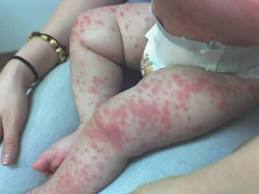
Symptoms can vary. It Must have a fever for more than five days, conjunctivitis, rash, changes in the oral mucosa, changes in the hands and feet, lack of effect of antibiotics, lymphadenopathy in the neck (often lacking).
Kawasaki disease prognosis

Since diagnosis is difficult to ask, many will get antibiotics without help.

The dilemma is the risk of complications and serious sequela.

Kawasaki syndrome

Kawasaki syndrome


Follow-up
Plan for follow-up depends on several factors. These include the age when one becomes ill, the effect of treatment and the size of any ballooning of the arteries. The current guidelines released in 2004.

Doctors found no or small, transient changes in a coronary arteries, there is no need for special attention beyond the medical checkups every 3 to five years. These controls should include an assessment of the overall risk of cardiovascular disease and counseling to reduce this. There is no need for special inspections or monitoring by the cardiologist.

Doctors found a small change in the coronary arteries; the child should continue with medication (ASA) until the changes have gone back. There are no restrictions on physical activity except for the first 6-8 weeks in children under 11 years. Children over 11 years of age must receive individual advice on physical activity depending on the findings on cardiac examinations. After the initial phase of illness, it is recommended annual follow-up by the cardiologist.
Kawasaki syndrome

In the case of larger or more ballooning of the arteries, put the child on long-term treatment with blood thinners. Contact Sports discouraged until the cardiologist may find that the changes have gone back. It is recommended follow-up twice a year by the cardiologist. The same recommendations apply if it is shown that the arteries are clogged.

Kawasaki disease treatment

Kawasaki disease treatment


Treatment
Since the condition is reminiscent of an infection and the diagnosis is difficult to ask, many will have received antibiotics without effect. Regardless of the disease in its acute phase transition by itself.

The dilemma is the risk of complications and serious sequela. If the diagnosis is made, doctors will therefore provide direct immunoglobulin in the blood to reduce the harmful effects of the disease. This treatment provides rapid improvement in the condition with fever of freedom within one to two days. The treatment also reduces the risk of later heart disease. There will also be given acetylsalicylic acid (ASA, ex. Dispril) in a few weeks.

Kawasaki disease diagnosis

How is the diagnosis?
Diagnosis is made primarily on the medical history, and finds the doctor do when the child is physically examined. Blood tests do not contribute to make any definite diagnosis, but may be instructive. X-ray of the lungs can sometimes show mild pneumonia-like changes.
Kawasaki disease treatment

If doctors suspect the diagnosis, the echocardiogram show changes in the heart that support the diagnosis.

Kawasaki syndrome symptoms

Kawasaki syndrome symptoms


What are the symptoms of Kawasaki syndrome?

Symptoms and signs develop over the first 10 days, and the disease goes back by itself in most children, even without treatment. Symptoms can vary, but most have:


  • Fever for more than five days without explanation
  • Conjunctivitis
  • Rash - are almost always.
  • Lymph node enlargement in the neck - often lack.
  • Changes in the oral mucosa
  • Changes at the hands and feet
  • Lack of effect of antibiotic

A number of other symptoms may also occur in connection with the disease: eg. irritability, joint pain, diarrhea, abdominal pain and ear pain. The changes in the oral cavity consist of redness, cracking and / or crust formation on the lips and strawberry tongue. The changes in hands and feet made of thickened skin, swelling and redness of the palms and soles, desquamation after approx. two weeks.
Kawasaki syndrome symptoms

Symptoms and signs develop over the first 10 days, and the disease goes back by itself in most children, even without treatment.

Kawasaki syndrome causes

Kawasaki syndrome causes


What causes Kawasaki Syndrome?
The cause is unknown. Probably, there are a number of factors that contribute to disease, but there are indications that there is an infection.

However, they have not been able to detect any virus or microbe.

In the acute phase developed inflammation of the wall of the coronary arteries (coronary vasculitis).

This can lead to a moderate expansion of the coronary artery.
Kawasaki syndrome causes

The lesions may persist from a few weeks to over a year after the disease onset.

What is Kawasaki Syndrome

What is Kawasaki Syndrome


What is Kawasaki Syndrome?

It is a febrile illness in children with rash, mucosal changes and lymph node enlargement. There is inflammation in the body's blood vessels (vasculitis). The condition is self-limiting and resolves by itself. Although the condition is by itself, one can get without late effects of treatment. The development of ballooning (aneurysm) of a coronary arteries (coronary arteries) in 20-25% if you do not receive treatment. In time, these sinuses interspersed cause serious complications. The disease was first described in Japan in the 1960s.

The disease affects children aged from three months to 12 years, 85% are under five years a . Isolated cases in adults are described. Sweden has found an annual incidence of 6.2 cases per 100,000 under five. The incidence of the disease varies throughout the year, with peaks in winter and spring.
What is Kawasaki Syndrome

The syndrome is most commonly in Asians, a little less frequently among blacks and lowest among whites. The incidence in the West is increasing.

Dilated cardiomyopathy prognosis

Dilated cardiomyopathy prognosis


Pregnancy-related cardiomyopathy

What is the prognosis?
Unlike the second cardiomyopathies are more people with cardiomyopathy periparital healthy by themselves, and they recover heart function. In a study from South Africa, 15% died, while 23% had recovered normal cardiac function after six months of treatment. Improvement, however, was observed both in the 2 and three year after the diagnosis was made, so that improvement phase is longer than the first 6-12 months.

A new pregnancy in a woman who has had periparital cardiomyopathy, provides a clearly increased risk of fresh heart failure. The greatest risk is for those who still have a remnant of heart failure after previous pregnancies.
Dilated cardiomyopathy prognosis

One study showed that half of those who became pregnant again, went through the next pregnancy, unless there were signs of new disease and heart failure.

Treatment cardiomyopathy

Treatment cardiomyopathy


Pregnancy-related cardiomyopathy

What is the treatment?
Since we do not know the cause of the disease, then we cannot give medicine to treat the disease itself. The treatment, therefore, be directed towards heart failure. Medications such as ACE inhibitors, beta blockers and digitalis use. Typical turnaround time is at least one year. In severe cases, it may be necessary with heart transplantation.

Otherwise you should be careful about using too much salt. You should also limit alcohol intake. It is important to try to be physically active.

How is the diagnosis?

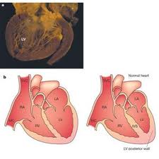
The diagnosis involves the exclusion of other possible explanations for the condition. The main investigation is ultrasound of the heart ( echocardiography ).
Treatment cardiomyopathy

This study can provide both descriptive pictures of the extent of heart muscle damage, and it can quantify the degree of eventual heart failure.

Signs and symptoms of cardiomyopathy

Signs and symptoms of cardiomyopathy


Pregnancy-related cardiomyopathy

What are the symptoms and signs, disease?
The condition usually presents with symptoms and signs of heart failure. There is an increased amount of water retention (swollen legs), breathing difficulty on exertion, breathing difficulty when lying but not when you sit up, nocturnal attacks of breathing difficulty, persistent cough, discomfort from the stomach, heart palpitations.

Early symptoms and signs are often overlooked and can be camouflaged by the pregnancy; they are perceived as normal pregnancy signs.


In some cases, thrombosis with obstruction of blood vessels (arteries) in the legs (trunde gangrene), brain (stroke), in the intestine (threatening gangrene), in the heart (heart attack). Such complications can be life threatening.